Arteries In Neck Labeled - 20.5 Circulatory Pathways - Anatomy & Physiology : At each level, the cervical vertebrae protect the spinal cord and work with muscles, tendons, ligaments, and joints to provide a combination of support, structure, and flexibility to the neck.
Arteries In Neck Labeled - 20.5 Circulatory Pathways - Anatomy & Physiology : At each level, the cervical vertebrae protect the spinal cord and work with muscles, tendons, ligaments, and joints to provide a combination of support, structure, and flexibility to the neck.. Branch of vertebral artery and thyrocervical trunk is labeled. May 31, 2021 · arteries of the cardiovascular system diagram practice test. Serious games to learn anatomy. Internal thoracic artery branches from same segment, but inferiorily, and is therefore not visible. These joints generally allow more movement than fibrous joints but less movement than synovial joints.
May 27, 2021 · the study of the vascularisation of the brain is possible with the arteries and venous sinuses sections. Download free books in pdf format. Superficial dissection of the right side of the neck, showing the carotid and subclavian arteries. Superior boundary (base) the lower border of the body of the mandible, and a line extending from the angle of the mandible to the mastoid process Anatomy ninja is an anatomy game based on medical illustrations, 3d images, radiographs and ct, mri images, that will help beginners as well as advanced players perfecting their lower and upper limb anatomy knowledge.
External carotid artery | Radiology Reference Article ... from prod-images-static.radiopaedia.org This feature has been chosen to show encephalic lobes: Pituitary and pineal glands are regrouped as "gland". Branch of vertebral artery and thyrocervical trunk is labeled. May 31, 2021 · arteries of the cardiovascular system diagram practice test. Read online books for free new release and bestseller Anatomy ninja is an anatomy game based on medical illustrations, 3d images, radiographs and ct, mri images, that will help beginners as well as advanced players perfecting their lower and upper limb anatomy knowledge. Cartilaginous joints are a type of joint where the bones are entirely joined by cartilage, either hyaline cartilage or fibrocartilage. As the cursor is moved over a particular anatomical area, that area is highlighted and labeled:
Cartilaginous joints are a type of joint where the bones are entirely joined by cartilage, either hyaline cartilage or fibrocartilage.
4,837 likes · 3 talking about this. Spend a few minutes analysing the diagram, and trying to connect the location of the structures with what you've learned in the video. Midline of the neck from chin to the jugular notch posterior boundary: This feature has been chosen to show encephalic lobes: Read online books for free new release and bestseller Superior boundary (base) the lower border of the body of the mandible, and a line extending from the angle of the mandible to the mastoid process In this diagram of the cardiovascular system, you can see labeled structures. Pituitary and pineal glands are regrouped as "gland". Serious games to learn anatomy. These joints generally allow more movement than fibrous joints but less movement than synovial joints. Seven cervical vertebrae, labeled c1 to c7, form the cervical spine from the base of the skull down to the top of the shoulders. The anterior margin of sternocleidomastoid: At each level, the cervical vertebrae protect the spinal cord and work with muscles, tendons, ligaments, and joints to provide a combination of support, structure, and flexibility to the neck.
Spend a few minutes analysing the diagram, and trying to connect the location of the structures with what you've learned in the video. Cartilaginous joints are a type of joint where the bones are entirely joined by cartilage, either hyaline cartilage or fibrocartilage. Superior boundary (base) the lower border of the body of the mandible, and a line extending from the angle of the mandible to the mastoid process Anatomy ninja is an anatomy game based on medical illustrations, 3d images, radiographs and ct, mri images, that will help beginners as well as advanced players perfecting their lower and upper limb anatomy knowledge. Pituitary and pineal glands are regrouped as "gland".
Top Photos in Blood supply of the neck from www.edoctoronline.com This feature has been chosen to show encephalic lobes: These joints generally allow more movement than fibrous joints but less movement than synovial joints. Midline of the neck from chin to the jugular notch posterior boundary: Download free books in pdf format. Seven cervical vertebrae, labeled c1 to c7, form the cervical spine from the base of the skull down to the top of the shoulders. Internal thoracic artery branches from same segment, but inferiorily, and is therefore not visible. Serious games to learn anatomy. Read online books for free new release and bestseller
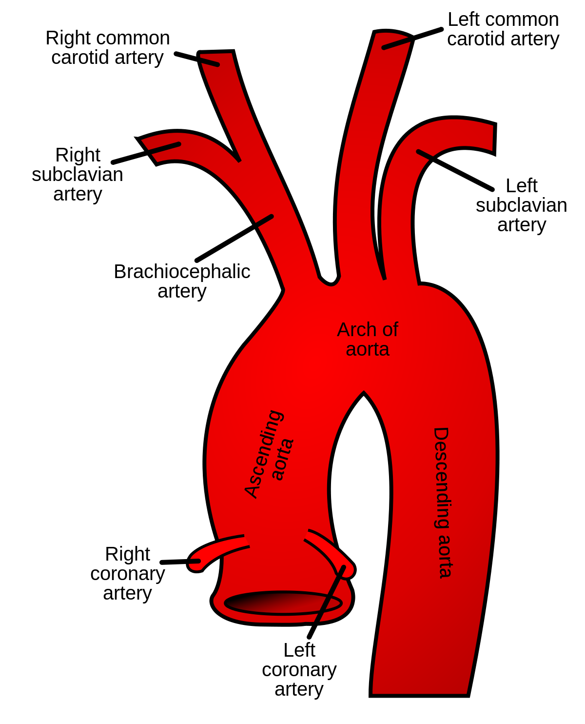
May 31, 2021 · arteries of the cardiovascular system diagram practice test.
Superficial dissection of the right side of the neck, showing the carotid and subclavian arteries. May 31, 2021 · arteries of the cardiovascular system diagram practice test. Superior boundary (base) the lower border of the body of the mandible, and a line extending from the angle of the mandible to the mastoid process Seven cervical vertebrae, labeled c1 to c7, form the cervical spine from the base of the skull down to the top of the shoulders. As the cursor is moved over a particular anatomical area, that area is highlighted and labeled: Serious games to learn anatomy. Pituitary and pineal glands are regrouped as "gland". Cartilaginous joints are a type of joint where the bones are entirely joined by cartilage, either hyaline cartilage or fibrocartilage. May 27, 2021 · the study of the vascularisation of the brain is possible with the arteries and venous sinuses sections. Spend a few minutes analysing the diagram, and trying to connect the location of the structures with what you've learned in the video. The anterior margin of sternocleidomastoid: These joints generally allow more movement than fibrous joints but less movement than synovial joints. In this diagram of the cardiovascular system, you can see labeled structures.
Spend a few minutes analysing the diagram, and trying to connect the location of the structures with what you've learned in the video. The anterior margin of sternocleidomastoid: Pituitary and pineal glands are regrouped as "gland". Anatomy ninja is an anatomy game based on medical illustrations, 3d images, radiographs and ct, mri images, that will help beginners as well as advanced players perfecting their lower and upper limb anatomy knowledge. Download free books in pdf format.
Common carotid artery - Wikipedia from upload.wikimedia.org Superior boundary (base) the lower border of the body of the mandible, and a line extending from the angle of the mandible to the mastoid process Branch of vertebral artery and thyrocervical trunk is labeled. Serious games to learn anatomy. Seven cervical vertebrae, labeled c1 to c7, form the cervical spine from the base of the skull down to the top of the shoulders. May 27, 2021 · the study of the vascularisation of the brain is possible with the arteries and venous sinuses sections. As the cursor is moved over a particular anatomical area, that area is highlighted and labeled: Midline of the neck from chin to the jugular notch posterior boundary: Anatomy ninja is an anatomy game based on medical illustrations, 3d images, radiographs and ct, mri images, that will help beginners as well as advanced players perfecting their lower and upper limb anatomy knowledge.
Read online books for free new release and bestseller
Midline of the neck from chin to the jugular notch posterior boundary: Cartilaginous joints are a type of joint where the bones are entirely joined by cartilage, either hyaline cartilage or fibrocartilage. Download free books in pdf format. May 31, 2021 · arteries of the cardiovascular system diagram practice test. At each level, the cervical vertebrae protect the spinal cord and work with muscles, tendons, ligaments, and joints to provide a combination of support, structure, and flexibility to the neck. Superficial dissection of the right side of the neck, showing the carotid and subclavian arteries. Branch of vertebral artery and thyrocervical trunk is labeled. 4,837 likes · 3 talking about this. As the cursor is moved over a particular anatomical area, that area is highlighted and labeled: These joints generally allow more movement than fibrous joints but less movement than synovial joints. This feature has been chosen to show encephalic lobes: The anterior margin of sternocleidomastoid: Pituitary and pineal glands are regrouped as "gland".
Seven cervical vertebrae, labeled c1 to c7, form the cervical spine from the base of the skull down to the top of the shoulders arteries in neck. The anterior margin of sternocleidomastoid:


Posting Komentar
0 Komentar